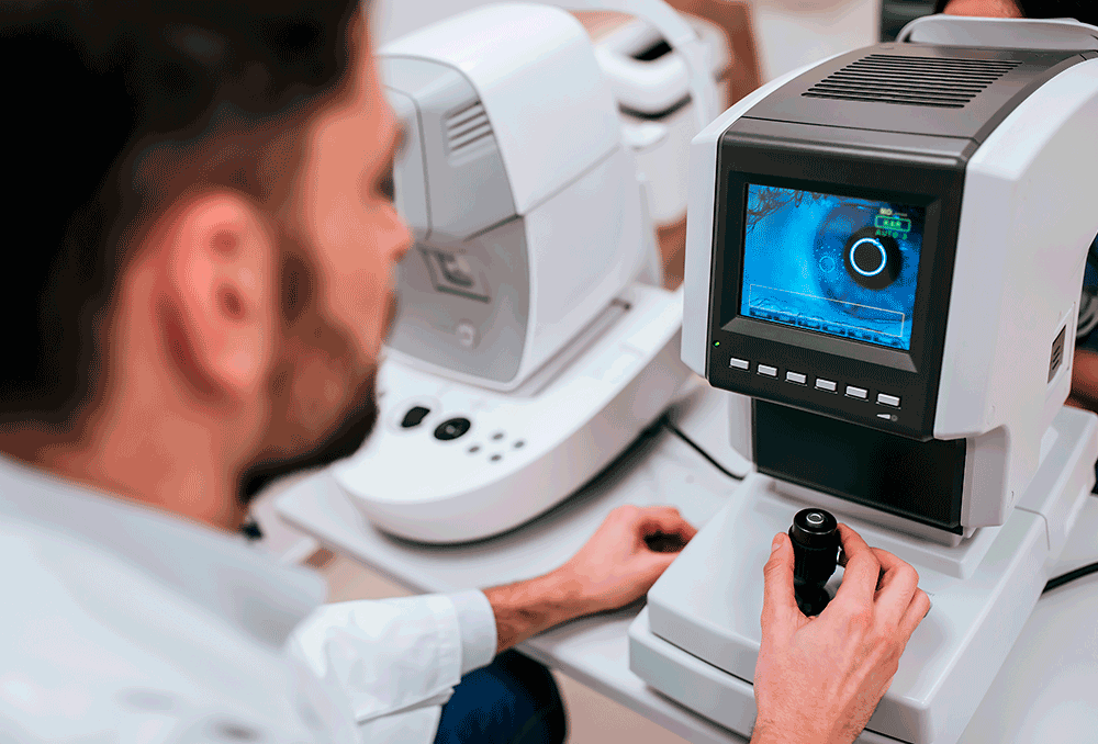
Ocular pachymetry is the examination which allows the thickness of the cornea to be measured, both in its central and more peripheral areas.
The cornea is a transparent structure without blood vessels, located in the most anterior part of the ocular globe, which is part of the eye's wall and works as a window, as it is completely transparent. A cornea is healthy when throughout its structure it does not present opacities, has a thickness within normal values, and has a curvature that allows images to be formed in a clear way on the retina.
In pachymetry, results are measured in microns or micras (thousandths of a mm). A central corneal thickness considered to be normal lies between 490 microns and 550 microns and in its peripheral zone, corneal thickness values can vary between 700 and 900 microns. Outside these normative values, in principle, changes may be causing thinning or increased corneal thickness. There are normal corneas outside these values, but in principle there is a pathology underlying these variations.
The reference method for measuring corneal thickness is still ultrasonic pachymetry because of its reliability and ease, it not only determines the central thickness of the cornea, but also the thickness at all desired points. It is a simple and quick examination. It is necessary to place anaesthetic drops of form (as the probe must be in contact with the surface of the cornea).
Other ways of measuring pachymetry are more convenient as there is no contact with the eye.
Corneal topography, by studying its entire structure, includes thickness measurement. One of the devices we use, the Pentacam, allows accurate maps to be obtained through 3D reconstruction of high-resolution photographs.
O OCT anterior segment OCT, more recent, allows even more precise images, achieving very accurate measurements.
Either method allows accurate measurement of corneal thickness.


